Feedback-01
feedback-01_x4934.2190_y68036.2190
HTT-TILS-001-03B.ndpi_x4934.2190_y68036.2190
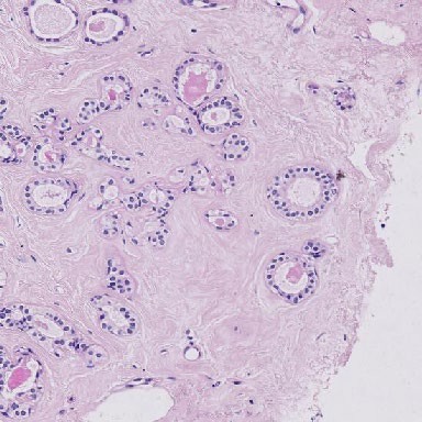
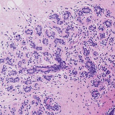
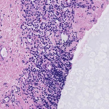
Expert Panel Annotations
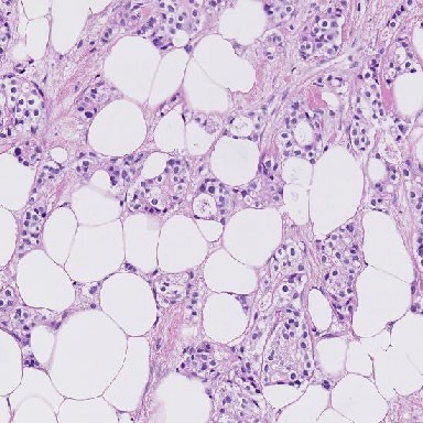
| ROI Type | Percent Tumor-Associated Stroma | sTILs Density |
|---|---|---|
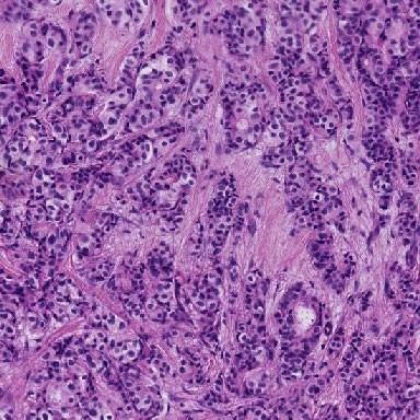
| Evaluable | 25 | 0 |
| Evaluable | 12 | 5 |
| Evaluable | 25 | 5 |
| Evaluable | 25 | 0 |
| Evaluable | 40 | 0 |
| Evaluable | 50 | 2 |
Mean Percent Tumor-Associated Stroma: 29.5
Mean sTILs Density: 2
Comments: Adipocytes are excluded from tumor-associated stroma.
Pitfalls: Adipocytes are not considered part of the tumor-associated stroma for purposes of sTILs assessment.
Feedback-02
feedback-02_x24343.2190_y11775.2190
HTT-TILS-001-04B.ndpi_x24343.2190_y11775.2190
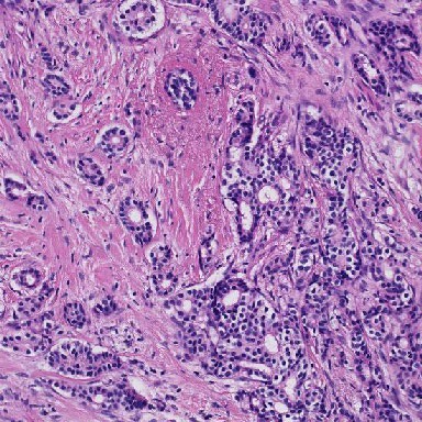
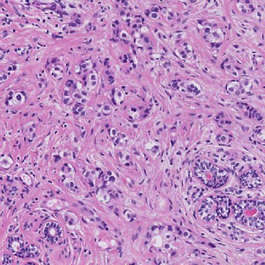
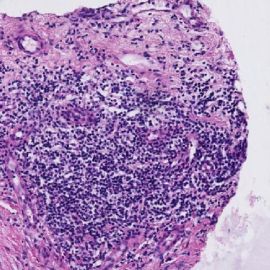
Expert Panel Annotations
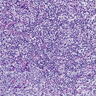
| ROI Type | Percent Tumor-Associated Stroma | sTILs Density |
|---|---|---|
| Evaluable | 30 | 90 |
| Evaluable | 60 | 95 |
| Evaluable | 50 | 92 |
| Evaluable | 50 | 75 |
| Evaluable | 60 | 90 |
| Evaluable | 60 | 90 |
Mean Percent Tumor-Associated Stroma: 51.7
Mean sTILs Density: 88.7
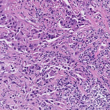
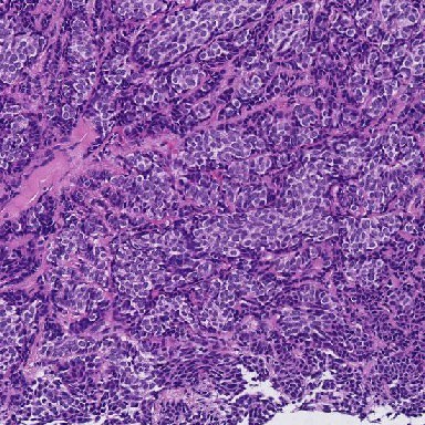
Comments: A challenging case. The high density of lymphocytes results in difficulty determining whether the lymphocytes are located in stroma, or whether they infiltrate tumor cell nests. The presence of small blood vessels and small gaps between lymphocytes suggest the lymphocytes reside within stroma. Occasional tumor cells with small nuclei (possibly degenerating) may be confused for lymphocytes.
Pitfalls: In regions where the sTILs density is very high, the underlying stroma may be obscured. Non-lymphocytes with small nuclei may be confused for lymphocytes.
Feedback-03
feedback-03_x29456.2190_y23838.2190
HTT-TILS-001-27B.ndpi_x29456.2190_y23838.2190
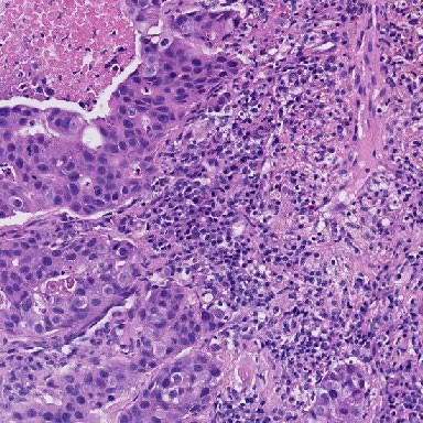
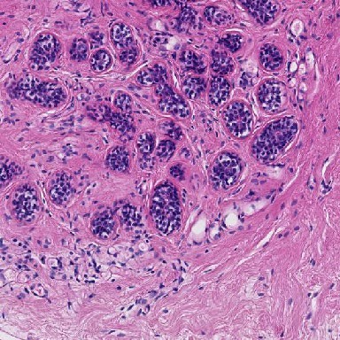
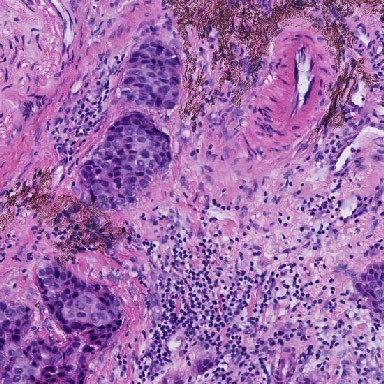
Expert Panel Annotations
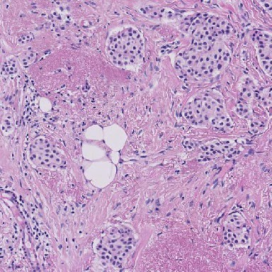
| ROI Type | Percent Tumor-Associated Stroma | sTILs Density |
|---|---|---|
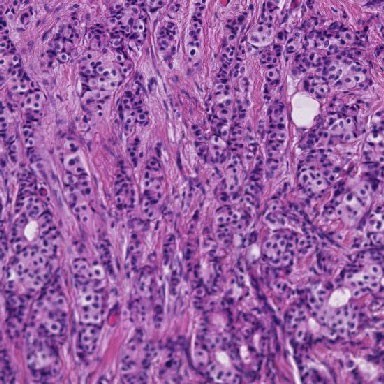
| Evaluable | 70 | 3 |
| Evaluable | 60 | 8 |
| Evaluable | 75 | 8 |
| Evaluable | 70 | 3 |
| Evaluable | 75 | 1 |
| Evaluable | 60 | 5 |
Mean Percent Tumor-Associated Stroma: 68.3
Mean sTILs Density: 4.7
Comments: There is clearly demarcated stroma in invasive tumor.
Pitfalls: Adipocytes are not considered part of the tumor-associated stroma for purposes of sTILs assessment. Areas of elastosis and fibroblastic proliferation are included as tumor-associated stroma.
Feedback-04
feedback-04_x5114.2190_y25709.2190
HTT-TILS-001-27B.ndpi_x5114.2190_y25709.2190
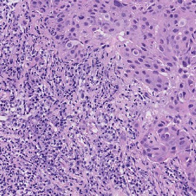
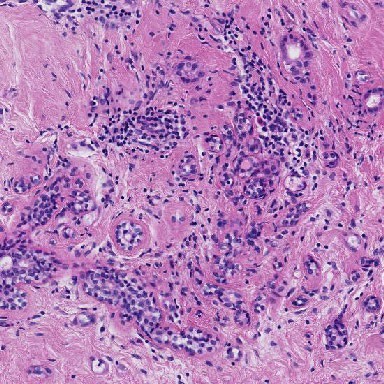
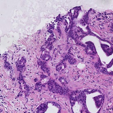
Expert Panel Annotations
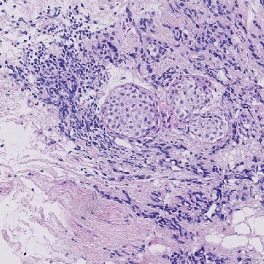
| ROI Type | Percent Tumor-Associated Stroma | sTILs Density |
|---|---|---|
| Evaluable | 60 | 15 |
| Evaluable | 8 | 20 |
| Evaluable | 75 | 13 |
| Evaluable | 70 | 3 |
| Evaluable | 80 | 10 |
| Evaluable | 80 | 10 |
Mean Percent Tumor-Associated Stroma: 62.2
Mean sTILs Density: 11.8
Comments: A challenging ROI. The DCIS in the center of the ROI is excluded from the calculation. Crushed tumor cells may be confused for lymphocytes. There are few small foci of invasive tumor (e.g. in upper third of ROI) that would be the focus of assessment. The lower part of ROI is also not reliable for scoring due to marked crush artifact. Crushed cells in the lower right quarter are suspicious for carcinoma, and not TILs, based on architecture similar to tumor cells outside of this ROI.
Pitfalls: DCIS is excluded from the numerator when calculating the percentage of tumor-associated stroma. Non-lymphocytes may be confused for lymphocytes if there is tissue fixation/preservation artifact.
Feedback-05
feedback-05_x12694.2190_y13922.2190
HTT-TILS-001-29B.ndpi_x12694.2190_y13922.2190
Expert Panel Annotations
| ROI Type | Percent Tumor-Associated Stroma | sTILs Density |
|---|---|---|
| Not Evaluable | NA | NA |
| Not Evaluable | NA | NA |
| Not Evaluable | NA | NA |
| Not Evaluable | NA | NA |
| Not Evaluable | NA | NA |
| Not Evaluable | NA | NA |
Mean Percent Tumor-Associated Stroma: NA
Mean sTILs Density: NA
Comments: A difficult case that would need further staining to confidently determine if could be tumor.
Pitfalls: Invasive carcinoma must be present to perform the sTILs assessment.
Feedback-06
feedback-06_x34191.2190_y65798.2190
HTT-TILS-001-32B.ndpi_x34191.2190_y65798.2190
Expert Panel Annotations
| ROI Type | Percent Tumor-Associated Stroma | sTILs Density |
|---|---|---|
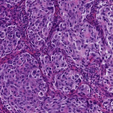
| Evaluable | 50 | 5 |
| Not Evaluable | NA | NA |
| Evaluable | 31 | 10 |
| Evaluable | 65 | 5 |
| Evaluable | 45 | 0 |
| Evaluable | 50 | 5 |
Mean Percent Tumor-Associated Stroma: 48.2
Mean sTILs Density: 5
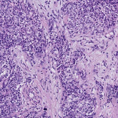
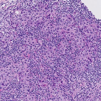
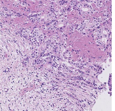
Comments: A subset of carcinoma cells exhibit perinuclear clearing/halos (e.g. 5 o’clock). Because these cells form cohesive nests and tubules, they can be recognized as carcinoma, and not lymphocytes. In addition, when assessing for percent tumor-associated stroma, exclude gland lumen from the area of tumor-associated stroma.
Pitfalls: Perinuclear clearing can cause challenges in discrimination of cells, such as macrophages, lymphocytes/plasma cells, tumor cells, or others. Additional immunohistochemical staining may be helpful to further subclassify such cells.
Feedback-07
feedback-07_x25888.2190_y33488.2190
HTT-TILS-001-33B.ndpi_x25888.2190_y33488.2190
Expert Panel Annotations
| ROI Type | Percent Tumor-Associated Stroma | sTILs Density |
|---|---|---|
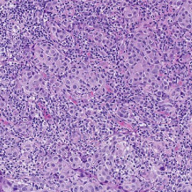
| Evaluable | 60 | 50 |
| Evaluable | 55 | 75 |
| Evaluable | 56 | 70 |
| Evaluable | 46 | 75 |
| Evaluable | 60 | 75 |
| Evaluable | 50 | 40 |
Mean Percent Tumor-Associated Stroma: 54.5
Mean sTILs Density: 64.2
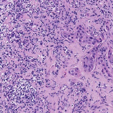
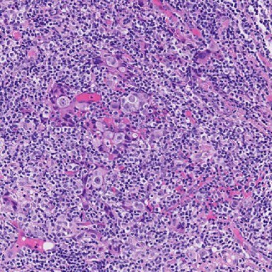
Comments: Around 2 o’clock, cells with perinuclear halos and eosinophilic cytoplasm could be plasma cells or macrophages. This ambiguity is due to the slide fixation, and they are likely all the same cell type; immunohistochemical staining could confirm their cell type(s). There was a considerable range of sTILs density scores among our experts; the 2 lower density scores of 40 and 50 may be a result of the perinuclear clearing in the 3 o’clock region centrally causing uncertainty as to whether those cells are macrophages or lymphocytes. There are also fibroblasts present that may be mistaken for TILs.
Pitfalls: Perinuclear clearing can cause challenges in discrimination of cells, such as macrophages, lymphocytes/plasma cells, tumor cells, or others. Additional immunohistochemical staining may be helpful to further subclassify such cells.
Feedback-08
feedback-08_x29652.2190_y6224.2190
HTT-TILS-001-33B.ndpi_x29652.2190_y6224.2190
Expert Panel Annotations
| ROI Type | Percent Tumor-Associated Stroma | sTILs Density |
|---|---|---|
| Evaluable | 70 | 85 |
| Evaluable | 60 | 70 |
| Evaluable | 50 | 75 |
| Evaluable | 70 | 85 |
| Evaluable | 60 | 80 |
| Evaluable | 60 | 60 |
Mean Percent Tumor-Associated Stroma: 61.7
Mean sTILs Density: 75.8
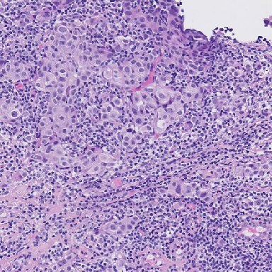
Comments: “This one’s easy in a sense, because the square is very nicely divided in triangles somewhat … it’s more than 50% of stroma and so it’s 55-60% stroma….” Despite perinuclear halos being prominent in this ROI, experts achieved better concordance; this is likely due to the arrangement of tumor and stroma in the ROI, where the tumor is all aggregated to one aspect.
Pitfalls: There are no significant pitfalls for this ROI.
Feedback-09
feedback-09_x29809.2190_y47938.2190 HTT-TILS-001-33B.ndpi_x29809.2190_y47938.2190
Expert Panel Annotations
| ROI Type | Percent Tumor-Associated Stroma | sTILs Density |
|---|---|---|
| Evaluable | 60 | 30 |
| Evaluable | 60 | 30 |
| Evaluable | 50 | 67 |
| Evaluable | 75 | 50 |
| Evaluable | 65 | 45 |
| Evaluable | 50 | 40 |
Mean Percent Tumor-Associated Stroma: 60
Mean sTILs Density: 43.7
Comments: This a suboptimal H&E fixation with lots of cellular degeneration. It has been demonstrated that poor staining and fixation has been associated with greater discordance among pathologist scores (Kos et al., npj Breast Cancer (2020)). This can lead to difficulty determining cell types, where immunohistochemical staining could confirm their cell type(s). Due to the problematic nature of this ROI, it may not be an ideal region to use in clinical practice.
Pitfalls: Perinuclear clearing can cause challenges in discrimination of cells, such as macrophages, lymphocytes/plasma cells, tumor cells, or others. Additional immunohistochemical staining may be helpful to further subclassify such cells.
Feedback-10
feedback-10_x32624.2190_y18730.2190 HTT-TILS-001-33B.ndpi_x32624.2190_y18730.2190
Expert Panel Annotations
| ROI Type | Percent Tumor-Associated Stroma | sTILs Density |
|---|---|---|
| Evaluable | 85 | 40 |
| Evaluable | 80 | 35 |
| Evaluable | 86 | 61 |
| Evaluable | 85 | 65 |
| Evaluable | 80 | 50 |
| Evaluable | 85 | 30 |
Mean Percent Tumor-Associated Stroma: 83.5
Mean sTILs Density: 46.8
Comments: The perinuclear clearing in the 6-9 o’clock region can cause difficulty distinguishing between macrophages and lymphocytes. Crush artifact (at 9 and 12 o’clock) may challenge the ability to categorize cells. Additional immunohistochemical staining could confirm their cell types(s). The uneven distribution of cell, especially the density differences between the right and left halves of the ROI, introduces additional uncertainty in determining an sTILs density for the entire ROI.
Pitfalls: Non-lymphocytes may be confused for lymphocytes if there is cellular preservation artifact.
Feedback-11
feedback-11_x13400.2190_y32583.2190 HTT-TILS-001-34B.ndpi_x13400.2190_y32583.2190
Expert Panel Annotations
| ROI Type | Percent Tumor-Associated Stroma | sTILs Density |
|---|---|---|
| Evaluable | 5 | 0 |
| Evaluable | 5 | 5 |
| Evaluable | 8 | 5 |
| Evaluable | 15 | 3 |
| Evaluable | 10 | 0 |
| Evaluable | 15 | 10 |
Mean Percent Tumor-Associated Stroma: 9.7
Mean sTILs Density: 3.8
Comments: Tumor cell eosinophilia is similar in appearance to stromal eosinophilia and causes difficulty in distinguishing these entities. The sTILs density can be inflated due to the edge artifact (upper part of lower zone) and crushed zone. An algorithm may recognize these as intraepithelial TILs since they are smaller than the cells in the upper part of the ROI.
Pitfalls: Occasionally, tumor cells may exhibit cytoplasm with eosinophilia similar to that of adjacent stroma, and thus be mistaken for stroma, potentially affecting the sTILs score. Non-lymphocytes may be confused for lymphocytes if there is tissue fixation artifact. Non-lymphocytes with small nuclei may be confused for lymphocytes.
Feedback-12
feedback-12_x2572.2190_y8345.2190
HTT-TILS-001-41B.ndpi_x2572.2190_y8345.2190
Expert Panel Annotations
| ROI Type | Percent Tumor-Associated Stroma | sTILs Density |
|---|---|---|
| Evaluable | 10 | 0 |
| Evaluable | 5 | 1 |
| Evaluable | 14 | 4 |
| Evaluable | 20 | 0 |
| Evaluable | 40 | 0 |
| Evaluable | 50 | 2 |
Mean Percent Tumor-Associated Stroma: 23.2
Mean sTILs Density: 1.2
Comments: Two pitfalls in assessing Percent Tumor-Associated Stroma are captured in this ROI. Adipocytes are excluded from tumoral stroma. In addition, there is empty space that one must remember to include when calculating the stromal percentage. One may try to mentally compress the stroma in one corner and visualize how much space it takes up of the entire ROI.
Pitfalls: Adipocytes are not considered part of tumor-associated stroma for purposes of sTILs assessment. The percent of tumor-associated-stromal is calculated with respect to the area of the entire ROI. Negative/empty space is in the total ROI area, the denominator of the Percent Tumor-Associated Stroma.
Feedback-13
feedback-13_x9321.2190_y18852.2190
HTT-TILS-001-42B.ndpi_x9321.2190_y18852.2190
Expert Panel Annotations
| ROI Type | Percent Tumor-Associated Stroma | sTILs Density |
|---|---|---|
| Evaluable | 30 | 5 |
| Evaluable | 40 | 9 |
| Evaluable | 50 | 7 |
| Evaluable | 50 | 3 |
| Evaluable | 40 | 1 |
| Evaluable | 50 | 5 |
Mean Percent Tumor-Associated Stroma: 43.3
Mean sTILs Density: 5
Comments: It is difficult to distinguish between fibroblasts and sTILs in this case. The cells in the middle of the ROI are a bit wider than the other cells, so they probably are cancer cells that have artifact as a result of tissue processing. Though strong suspicion for a cancer cell, it could be a macrophage, which we see after treatment, and expect that an algorithm will have difficulty making this distinction on H&E stain.
Pitfalls: Non-lymphocytes may be confused for lymphocytes if there is tissue fixation artifact. Axially sectioned fibroblasts may be mistaken for lymphocytes.
Feedback-14
feedback-14_x39884.2190_y27444.2190 HTT-TILS-001-50B.ndpi_x39884.2190_y27444.2190
Expert Panel Annotations
| ROI Type | Percent Tumor-Associated Stroma | sTILs Density |
|---|---|---|
| Not Evaluable | NA | NA |
| Evaluable | 15 | 5 |
| Evaluable | 15 | 4 |
| Evaluable | 10 | 5 |
| Evaluable | 20 | 1 |
| Evaluable | 15 | 1 |
Mean Percent Tumor-Associated Stroma: 15
Mean sTILs Density: 3.2
Comments: This intra-tumoral stroma case is a “good case because cells are very pink, as much as pink as the stroma in between. So, it’s not so easy to identify the stroma which is in between the cancer cells.” Looking between three and six o’clock, this region is harder to assess than those in the upper right because you can’t see the cell borders very clearly.
Pitfalls: Occasionally, tumor cells may exhibit cytoplasm with eosinophilia similar to that of adjacent stroma, and thus be mistaken for stroma, potentially affecting the sTILs score.
Feedback-15
feedback-15_x22110.2190_y13641.2190
HTT-TILS-001-52B.ndpi_x22110.2190_y13641.2190
Expert Panel Annotations
| ROI Type | Percent Tumor-Associated Stroma | sTILs Density |
|---|---|---|
| Evaluable | 15 | 2 |
| Evaluable | 5 | 1 |
| Evaluable | 13 | 4 |
| Evaluable | 25 | 2 |
| Evaluable | 15 | 0 |
| Evaluable | 10 | 10 |
Mean Percent Tumor-Associated Stroma: 13.8
Mean sTILs Density: 3.2
Comments: There are no additional comments.
Pitfalls: There are no significant pitfalls for this ROI.
Feedback-16
feedback-16_x71829.2190_y49452.2190 HTT-TILS-001-52B.ndpi_x71829.2190_y49452.2190
Expert Panel Annotations
| ROI Type | Percent Tumor-Associated Stroma | sTILs Density |
|---|---|---|
| Evaluable | 15 | 0 |
| Evaluable | 5 | 0 |
| Evaluable | 11 | 5 |
| Evaluable | 20 | 0 |
| Evaluable | 20 | 0 |
| Evaluable | 15 | 1 |
Mean Percent Tumor-Associated Stroma: 14.3
Mean sTILs Density: 1
Comments: Here, we see the invasive margin with 10% stroma and very sTILs. From an expert panelist, “this is actually a good case because all these very tiny dark round things, small round blue cells, are just probably a fixation artifact of ischemic tumor cells, and these are easily overlooked by machine learning process as being TILs.” At 8 o’clock, those dark cells are more likely tumor cells than lymphocytes and may be the result of preservation and/or staining artifacts.
Pitfalls: Non-lymphocytes may be confused for lymphocytes if there is tissue fixation artifact.
Feedback-17
feedback-17_x28888.2190_y28271.2190 HTT-TILS-001-53B.ndpi_x28888.2190_y28271.2190

Expert Panel Annotations
| ROI Type | Percent Tumor-Associated Stroma | sTILs Density |
|---|---|---|
| Not Evaluable | NA | NA |
| Not Evaluable | NA | NA |
| Not Evaluable | NA | NA |
| Not Evaluable | NA | NA |
| Not Evaluable | NA | NA |
| Not Evaluable | NA | NA |
Mean Percent Tumor-Associated Stroma: NA
Mean sTILs Density: NA
Comments: There are no tumor cells within the thumbnail of the ROI. Outside of the ROI beyond the lower right corner, we see fibrotic tissue and ducts, but it is unclear is there are tumor cells here.
Pitfalls: Invasive carcinoma must be present for sTILs density assessment.
Feedback-18
feedback-18_x12938.2190_y7977.2190 HTT-TILS-001-57B.ndpi_x12938.2190_y7977.2190
Expert Panel Annotations
| ROI Type | Percent Tumor-Associated Stroma | sTILs Density |
|---|---|---|
| Evaluable | 30 | 0 |
| Evaluable | 20 | 1 |
| Evaluable | 25 | 0 |
| Evaluable | 40 | 3 |
| Evaluable | 40 | 0 |
| Evaluable | 40 | 2 |
Mean Percent Tumor-Associated Stroma: 32.5
Mean sTILs Density: 1
Comments: There are no additional comments.
Pitfalls: There are no significant pitfalls for this ROI.
Feedback-19
feedback-19_x16512.2190_y8168.2190 HTT-TILS-001-57B.ndpi_x16512.2190_y8168.2190
Expert Panel Annotations
| ROI Type | Percent Tumor-Associated Stroma | sTILs Density |
|---|---|---|
| Evaluable | 20 | 0 |
| Evaluable | 25 | 0 |
| Evaluable | 21 | 0 |
| Evaluable | 45 | 0 |
| Evaluable | 35 | 0 |
| Evaluable | 40 | 1 |
Mean Percent Tumor-Associated Stroma: 31
Mean sTILs Density: 0.2
Comments: There will be areas of out-of-focus because there are different thicknesses across the whole region.
Pitfalls: There will be areas of out-of-focus because there are different thicknesses across the whole region, or bad scan quality can contribute to an inaccurate assessment of TILs.
Feedback-20
feedback-20_x9143.2190_y47362.2190 HTT-TILS-001-73B.ndpi_x9143.2190_y47362.2190
Expert Panel Annotations
| ROI Type | Percent Tumor-Associated Stroma | sTILs Density |
|---|---|---|
| Evaluable | 10 | 50 |
| Evaluable | 10 | 80 |
| Evaluable | 13 | 59 |
| Evaluable | 75 | 50 |
| Evaluable | 35 | 60 |
| Evaluable | 10 | 30 |
Mean Percent Tumor-Associated Stroma: 25.5
Mean sTILs Density: 54.8
Comments: In this slide, we have a dark H&E stain and can see about 5 axes of fibrous stroma in the ROI with 3 of them not having sTILs. The axes of fibrous stroma are challenging because it is unclear if this is all to be considered stroma, such as capillaries or other poorly visualized structures in the band. When there is not a lot of stroma present, it becomes harder to distinguish the density of TILs, but it is not likely to have the high density estimate of 80 sTILs in this ROI. One of the dogmas of sTILs scoring is that it is not required for lymphocytes to have the same density as in a lymph node. It is okay for there to be space in between the lymphocytes.
Pitfalls: There are no significant pitfalls for this ROI.
Feedback-21
feedback-21_x15338.2190_y44740.2190 HTT-TILS-001-76B.ndpi_x15338.2190_y44740.2190
Expert Panel Annotations
| ROI Type | Percent Tumor-Associated Stroma | sTILs Density |
|---|---|---|
| Evaluable | 50 | 95 |
| Evaluable | 35 | 95 |
| Evaluable | 47 | 92 |
| Evaluable | 50 | 95 |
| Evaluable | 40 | 90 |
| Evaluable | 40 | 95 |
Mean Percent Tumor-Associated Stroma: 43.7
Mean sTILs Density: 93.7
Comments: There are no additional comments.
Pitfalls: There are no significant pitfalls for this ROI.
Feedback-22
feedback-22_x16170.2190_y42769.2190 HTT-TILS-001-76B.ndpi_x16170.2190_y42769.2190
Expert Panel Annotations
| ROI Type | Percent Tumor-Associated Stroma | sTILs Density |
|---|---|---|
| Evaluable | 40 | 90 |
| Evaluable | 25 | 95 |
| Evaluable | 50 | 86 |
| Evaluable | 40 | 95 |
| Evaluable | 30 | 90 |
| Evaluable | 30 | 95 |
Mean Percent Tumor-Associated Stroma: 35.8
Mean sTILs Density: 91.8
Comments: When assessing sTILs, remember that the sTILs percentage is in proportion to the area of the tumor-associated stroma, not tumor cellularity or cell count percentage which will give you a different score. In sTILs assessment, the denominator is the area of tumor-associated stroma in the given ROI, and the numerator is the area of sTILs in that tumor-associated stroma.
Pitfalls: The percent of tumor-associated-stromal is calculated with respect to the area of the entire ROI. Negative/empty space is in the total ROI area, the denominator of the Percent Tumor-Associated Stroma.
Feedback-23
feedback-23_x24500.2190_y76408.2190 HTT-TILS-001-76B.ndpi_x24500.2190_y76408.2190
Expert Panel Annotations
| ROI Type | Percent Tumor-Associated Stroma | sTILs Density |
|---|---|---|
| Evaluable | 90 | 90 |
| Evaluable | 60 | 95 |
| Evaluable | 75 | 95 |
| Evaluable | 75 | 95 |
| Evaluable | 80 | 90 |
| Evaluable | 80 | 90 |
Mean Percent Tumor-Associated Stroma: 76.7
Mean sTILs Density: 92.5
Comments: This is intra-tumoral stroma with high stromal and sTILs densities.
Pitfalls: Perinuclear clearing can cause challenges in discrimination of cells, such as macrophages, lymphocytes/plasma cells, tumor cells, or others. Additional immunohistochemical staining may be helpful to further subclassify such cells. In regions where the sTILs density is very high, the underlying stroma may be obscured.
Feedback-24
feedback-24_x29068.2190_y50307.2190 HTT-TILS-001-76B.ndpi_x29068.2190_y50307.2190
Expert Panel Annotations
| ROI Type | Percent Tumor-Associated Stroma | sTILs Density |
|---|---|---|
| Evaluable | 60 | 70 |
| Evaluable | 45 | 90 |
| Evaluable | 67 | 75 |
| Evaluable | 60 | 85 |
| Evaluable | 60 | 85 |
| Evaluable | 50 | 70 |
Mean Percent Tumor-Associated Stroma: 57
Mean sTILs Density: 79.2
Comments: There are 2 classical pitfalls demonstrated: crush zones (3 o’clock) and possible apoptotic tumor cells. Crushed cells become very dark and small, while apoptotic cells are round, blue cells. Both of which, if misinterpreted, can inflate the sTILs count. Fixation artifacts also give you this type of very small, once dark nuclei.
Pitfalls: Degenerating non-lymphocytes (e.g. pyknotic tumor cells) may be mistaken for lymphocytes. Non-lymphocytes may be confused for lymphocytes if there is tissue fixation and/or cellular preservation artifact. Perinuclear clearing can cause challenges in discrimination of cells, such as macrophages, lymphocytes/plasma cells, tumor cells, or others. Additional immunohistochemical staining may be helpful to further subclassify such cells. In regions where the sTILs density is very high, the underlying stroma may be obscured.
Feedback-25
feedback-25_x28631.2190_y33655.2190 HTT-TILS-001-80B.ndpi_x28631.2190_y33655.2190
Expert Panel Annotations
| ROI Type | Percent Tumor-Associated Stroma | sTILs Density |
|---|---|---|
| Evaluable | 90 | 90 |
| Evaluable | 90 | 90 |
| Not Evaluable | NA | NA |
| Not Evaluable | NA | NA |
| Evaluable | 99 | 0 |
| Not Evaluable | NA | NA |
Mean Percent Tumor-Associated Stroma: 93
Mean sTILs Density: 60
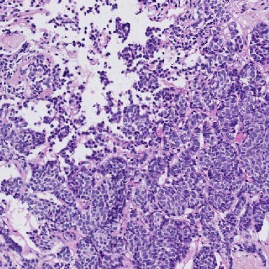
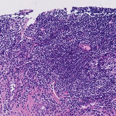
Comments: A challenging case that highlights the limitations of core biopsies. It may appear that we are at the edge of tumor though we don’t see an invasive margin. There is DCIS present to the right of this ROI, which should not be considered in sTILs evaluation. After discussion, group consensus was that this ROI is “Not Evaluable” for sTILs. This ROI might actually represent a lymphoid aggregate, which is not evaluated for sTILs. We suspect this is an aggregate because there is no stroma visualized between the TILs. Making the distinction between a lymphoid aggregate and stromal TILs is a challenging task for AI/ML algorithms.
Pitfalls: DCIS should be excluded from the numerator when calculating the percentage of tumor-associated stroma. Invasive carcinoma must be present to perform the sTILs assessment.
Feedback-26
feedback-26_x52173.2190_y57416.2190 HTT-TILS-001-80B.ndpi_x52173.2190_y57416.2190
Expert Panel Annotations
| ROI Type | Percent Tumor-Associated Stroma | sTILs Density |
|---|---|---|
| Not Evaluable | NA | NA |
| Not Evaluable | NA | NA |
| Not Evaluable | NA | NA |
| Not Evaluable | NA | NA |
| Not Evaluable | NA | NA |
| Not Evaluable | NA | NA |
Mean Percent Tumor-Associated Stroma: NA
Mean sTILs Density: NA
Comments: This is normal tissue.
Pitfalls: Invasive carcinoma must be present to perform the sTILs assessment.
Feedback-27
feedback-27_x56330.2190_y11332.2190 HTT-TILS-001-80B.ndpi_x56330.2190_y11332.2190
Expert Panel Annotations
| ROI Type | Percent Tumor-Associated Stroma | sTILs Density |
|---|---|---|
| Evaluable | 53 | 3 |
| Evaluable | 70 | 10 |
| Evaluable | 60 | 10 |
| Evaluable | 60 | 0 |
| Evaluable | 70 | 3 |
| Evaluable | 80 | 3 |
Mean Percent Tumor-Associated Stroma: 65.5
Mean sTILs Density: 4.8
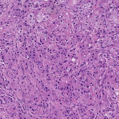
Comments: In the center of this ROI, we can see fibroblasts that have been transversely sliced, which can be mistaken as lymphocytes. Most of the lymphocytes can be found in the upper left corner and fibroblasts can be seen in the 3 o’clock position where the fibroblasts appear slightly more elongated. Invasive carcinoma can be seen at the 9 o’clock and 10:30 positions with normal glands in bottom left (7 o’clock) and right (5 o’clock) corners.
Pitfalls: Axially sectioned fibroblasts may be mistaken for lymphocytes. DCIS and normal glands should be excluded from the numerator when calculating the percentage of tumor-associated stroma.
Feedback-28
feedback-28_x57261.2190_y59992.2190 HTT-TILS-001-80B.ndpi_x57261.2190_y59992.2190
Expert Panel Annotations
| ROI Type | Percent Tumor-Associated Stroma | sTILs Density |
|---|---|---|
| Not Evaluable | NA | NA |
| Not Evaluable | NA | NA |
| Not Evaluable | NA | NA |
| Not Evaluable | NA | NA |
| Not Evaluable | NA | NA |
| Not Evaluable | NA | NA |
Mean Percent Tumor-Associated Stroma: NA
Mean sTILs Density: NA
Comments: This is normal tissue.
Pitfalls: Invasive carcinoma must be present to perform the sTILs assessment.
Feedback-29
feedback-29_x17750.2190_y20193.2190 HTT-TILS-001-83B.ndpi_x17750.2190_y20193.2190
Expert Panel Annotations
| ROI Type | Percent Tumor-Associated Stroma | sTILs Density |
|---|---|---|
| Not Evaluable | NA | NA |
| Not Evaluable | NA | NA |
| Not Evaluable | NA | NA |
| Evaluable | 75 | 5 |
| Not Evaluable | NA | NA |
| Not Evaluable | NA | NA |
Mean Percent Tumor-Associated Stroma: 75
Mean sTILs Density: 5
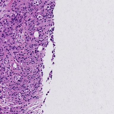
Comments: There are only 3 or so abnormal glands in this view that represent infiltrating tumor. However, it is expected to see lymphocytes in this ROI given the extent of normal tissue and scant tumor presence overall. This would not be an ROI to evaluate for stromal density. Any sTILs in this slide would have to be limited to the peri-tumoral stromal region which is minimal.
Pitfalls: Invasive carcinoma must be present to perform the sTILs assessment.
Feedback-30
feedback-30_x24381.2190_y16599.2190 HTT-TILS-001-83B.ndpi_x24381.2190_y16599.2190
Expert Panel Annotations
| ROI Type | Percent Tumor-Associated Stroma | sTILs Density |
|---|---|---|
| Not Evaluable | NA | NA |
| Not Evaluable | NA | NA |
| Not Evaluable | NA | NA |
| Evaluable | 56 | 0 |
| Not Evaluable | NA | NA |
| Not Evaluable | NA | NA |
Mean Percent Tumor-Associated Stroma: 56
Mean sTILs Density: 0
Comments: This is not tumor tissue.
Pitfalls: Invasive carcinoma must be present to perform the sTILs assessment.
Feedback-31
feedback-31_x9416.2190_y29452.2190 HTT-TILS-001-84B.ndpi_x9416.2190_y29452.2190
Expert Panel Annotations
| ROI Type | Percent Tumor-Associated Stroma | sTILs Density |
|---|---|---|
| Not Evaluable | NA | NA |
| Not Evaluable | NA | NA |
| Not Evaluable | NA | NA |
| Not Evaluable | NA | NA |
| Not Evaluable | NA | NA |
| Not Evaluable | NA | NA |
Mean Percent Tumor-Associated Stroma: NA
Mean sTILs Density: NA
Comments: No invasive tumor cells present or adjacent to ROI.
Pitfalls: Invasive carcinoma must be present to perform the sTILs assessment.
Feedback-32
feedback-32_x20669.2190_y12026.2190 HTT-TILS-001-86B.ndpi_x20669.2190_y12026.2190
Expert Panel Annotations
| ROI Type | Percent Tumor-Associated Stroma | sTILs Density |
|---|---|---|
| Evaluable | 75 | 30 |
| Evaluable | 35 | 60 |
| Evaluable | 86 | 15 |
| Evaluable | 75 | 30 |
| Evaluable | 70 | 25 |
| Evaluable | 70 | 20 |
Mean Percent Tumor-Associated Stroma: 68.5
Mean sTILs Density: 30
Comments: There was high variability among our expert panel. The challenge in this case is the handling of the thick-walled vessel, determining if perivascular infiltration is present, and uncertainty of the structure in the upper left-hand corner of the ROI. This may present a challenge to pathologists when assessing sTILs in an ROI-based manner because the vessel would not have been considered when investigating a slide digitally or on a microscope. However, for a machine learning tool, it is important to define. Similar to infiltrating immune cells in adipose tissue, if there is no mesenchymal fibroblasts in between there is not where for immune cells can bind to. “If we consider that reasoning that you don’t see TILs binding into the into the media of this vessel, we exclude it.”
Pitfalls: The walls of thick-walled vessels are not included as part of tumor-associated stroma.
Feedback-33
feedback-33_x38756.2190_y32439.2190 HTT-TILS-001-88B.ndpi_x38756.2190_y32439.2190

Expert Panel Annotations
| ROI Type | Percent Tumor-Associated Stroma | sTILs Density |
|---|---|---|
| Not Evaluable | NA | NA |
| Not Evaluable | NA | NA |
| Evaluable | 4 | 0 |
| Not Evaluable | NA | NA |
| Evaluable | 10 | 0 |
| Evaluable | 20 | 1 |
Mean Percent Tumor-Associated Stroma: 11.3
Mean sTILs Density: 0.3
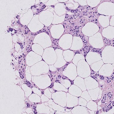
Comments: This ROI highlights the importance of understanding how evaluable stroma is defined. Though adipocytes may have biological significance in some scenarios, for the purpose of sTILs assessments, we don’t consider them as tumor-associated stroma. Therefore, the amount of stroma between the adipose tissue in this ROI is low. Given the low-grade atypia, one may confuse the cancer cells as lymphocyte nuclei because they are very small and dark, but one may find a lymphocyte in the top right region of the ROI.
Pitfalls: Adipocytes are not considered part of the tumor-associated stroma for purposes of sTILs assessment. Non-lymphocytes with small nuclei may be confused for lymphocytes.
Feedback-34
feedback-34_x16220.2190_y7255.2190 HTT-TILS-001-89B.ndpi_x16220.2190_y7255.2190
Expert Panel Annotations
| ROI Type | Percent Tumor-Associated Stroma | sTILs Density |
|---|---|---|
| Not Evaluable | NA | NA |
| Not Evaluable | NA | NA |
| Evaluable | 33 | 8 |
| Evaluable | 50 | 5 |
| Evaluable | 60 | 1 |
| Evaluable | 50 | 5 |
Mean Percent Tumor-Associated Stroma: 48.2
Mean sTILs Density: 4.8
Comments: Outside of the ROI, to the lower right, the tissue appears as invasive margin while DCIS can also be seen in the upper portion of the ROI. DCIS should not influence or be considered in the assessment of stromal percentage. In the upper right and 5 o’clock regions, the sTILs determination can be challenging due to possible perpendicularly cut fibroblasts. Another important consideration is the empty spacy must be considered when calculating the percent of tumor-associated stroma. Stromal percentage is calculated with respect to the area of the ROI and not with respect to the amount of tumor tissue present. In the clinical setting, sTILs assessment is centered on the proportion of sTILs to tumor-associated stroma; the calculation of percentage of tumor-associated stroma is not performed. For the development of a reference standard for machine learning algorithms, the algorithm will calculate the percentage of tumor-associated stroma within a given ROI as part of its analysis. Percent Tumor-Associated Stroma is relative to the square, not the tissue.
Pitfalls: Axially sectioned fibroblasts may be mistaken for lymphocytes. DCIS should be excluded from the numerator when calculating the percentage of tumor-associated stroma. The Percent Tumor-Associated Stroma is calculated with respect to the area of the entire ROI. Negative/empty space is to be included in the total ROI area, the denominator of the Percent Tumor-Associated Stroma.
Feedback-35
feedback-35_x12601.2190_y19050.2190 HTT-TILS-001-43B.ndpi_x12601.2190_y19050.2190
Expert Panel Annotations
| ROI Type | Percent Tumor-Associated Stroma | sTILs Density |
|---|---|---|
| Evaluable | 65 | 5 |
| Evaluable | 70 | 15 |
| Evaluable | 65 | 10 |
| Evaluable | 30 | 20 |
| Evaluable | 40 | 10 |
| Evaluable | 60 | 5 |
Mean Percent Tumor-Associated Stroma: 55
Mean sTILs Density: 10.8
Comments: Stroma with hyalinization and elastosis are included, as long as invasive cancer cells are present, in the TIL assessment. Necrotic tissue is excluded.
Pitfalls: Necrosis is excluded from sTILs evaluation. Hyalinized and elastotic stroma are included in assessment if associated with invasive carcinoma.
Feedback-36
feedback-36_x20589.2190_y28549.2190 HTT-TILS-001-50B.ndpi_x20589.2190_y28549.2190
Expert Panel Annotations
| ROI Type | Percent Tumor-Associated Stroma | sTILs Density |
|---|---|---|
| Evaluable | 35 | 30 |
| Evaluable | 50 | 20 |
| Evaluable | 35 | 20 |
| Evaluable | 35 | 20 |
| Evaluable | 31 | 41 |
| Evaluable | 40 | 25 |
Mean Percent Tumor-Associated Stroma: 37.7
Mean sTILs Density: 26
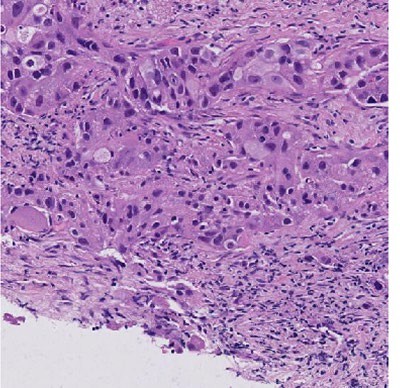
Comments: Crushed cells are difficult to assess in TILs, which are found in the lower-right hand corner between 3-6 o’clock. AI/ML algorithms may find it difficult to identify crushed lymphocytes.
Pitfalls: The Percent Tumor-Associated Stroma is calculated with respect to the area of the entire ROI. Negative/empty space is to be included in the total ROI area, the denominator or Percent Tumor-Associated Stroma. Non-lymphocytes may be confused for lymphocytes if there is cellular preservation artifact.